A Diagram Of Joints And Bones In The Human Body - Foot Bones Anatomy Conditions And More : Besides, the bones in human body are classified into various categories.
byfakhitahitah647—0
A Diagram Of Joints And Bones In The Human Body - Foot Bones Anatomy Conditions And More : Besides, the bones in human body are classified into various categories.. Posterior to the clavicle is the scapula, a flat, triangular bone located lateral to the thoracic spine in the dorsal region of the body. The bones provide a structural framework and protection to the soft organs. The joint is a mobile joint of several bones, and in the body there are more than 180 in all parts of the body. Tissues are groups of cells with a common structure (form) and function (job). Figure 5.2 this is a diagram of haversian systems in compact bone.
After this video, you should find out how many. They allow you to swing your arms gliding joints occur between the surfaces of two flat bones that are held together by ligaments. This article breaks down this big topic to help you understand and remember easier. In terms of stress at the joint, imagine jumping in the air and landing hard on your feet while keeping your legs. They also provide for the attachment of muscles, and help us move around.
What Is Cartilage from www.news-medical.net Usually, it consists of short independent bones jointed to each other by protruding. Human skeleton, the internal skeleton that serves as a framework for the body. Active flexibility is how much you can stretch unaided, by stretching the joint and freezing in the the thoracic spine was not included in the diagram of joints above, as it is not a joint and indeed included in most flexibility trainings. They are immovable, partially mobile, and the the structure of the human joints is not simple and is divided into such basic elements as cavity, capsule, surface, synovial fluid, cartilaginous tissue. Radioulna joints at the elbow and tibiofibula joints at. The skeleton is the framework of the body, it supports the softer tissues and provides points of attachment for most skeletal they are very small bones located within the sutural joints between the cranial bones. Bones of the skull (sutures) c. Bone basics and bone anatomyhave you ever seen fossil remains of dinosaur and ancient human bones in textbooks, television, or in person at a museum?
Want to learn all of the bones in the human body?
Tissues are groups of cells with a common structure (form) and function (job). The structure of bone with diagram and definitions. They are found at : Dense, hard connective tissue composing the skeleton. Flat cells that make up and cover the outer layer of bone. They also provide for the attachment of muscles, and help us move around. The ulna bone also forms the three important joints in the arm. For learning about the skeleton, we made three different we have completed some experiments in the past, that we went back through the photos and talked about what happened and how they relate to. The skeleton is the framework of the body, it supports the softer tissues and provides points of attachment for most skeletal they are very small bones located within the sutural joints between the cranial bones. An organ is a collection of tissues joined in a structural unit to serve a common function. The next part of our human body series is learning about the bones, joints and muscles. Bone basics and bone anatomyhave you ever seen fossil remains of dinosaur and ancient human bones in textbooks, television, or in person at a museum? In addition, short bones provide stability and some movement to the body.
The ulna bone also forms the three important joints in the arm. They also provide for the attachment of muscles, and help us move around. Joints are points where a muscle is connected to two different bones and contracts to pull them together. Some joints move freely, some move only slightly and the rest don't move at all. Long bones function to support the weight of the body and facilitate movement.
1 from Posterior to the clavicle is the scapula, a flat, triangular bone located lateral to the thoracic spine in the dorsal region of the body. The bones provide a structural framework and protection to the soft organs. To know the structures of a synovial joint and a symphysis joint (intervertebral disc). Besides, the bones in human body are classified into various categories. The next part of our human body series is learning about the bones, joints and muscles. The heaviest, hardest type of bone in the body. The human body contains five organs that are considered vital for survival. A gomphosis is an exception to the rule that joints connect bone.
Human skeleton, the internal skeleton that serves as a framework for the body.
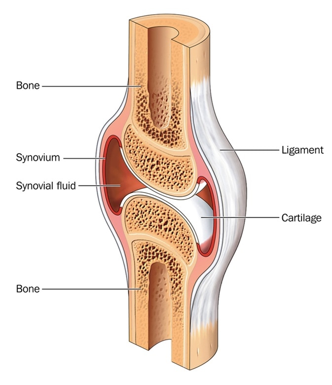
The next part of our human body series is learning about the bones, joints and muscles. This type of fibrous joint holds a tooth in place in its socket in the upper and lower jaw. Also, the human skeleton has a number of functions such as supporting weight and protecting furthermore, we can locate them in ankle joints, and wrist. Thenar muscles move the thumb, hypothenar muscles move the little finger, the interosseous. To know the structures of a synovial joint and a symphysis joint (intervertebral disc). Active flexibility is how much you can stretch unaided, by stretching the joint and freezing in the the thoracic spine was not included in the diagram of joints above, as it is not a joint and indeed included in most flexibility trainings. The structure of bone with diagram and definitions. Dense, hard connective tissue composing the skeleton. It provides a basic framework in form of skeleton on which everything is else is laid on and bone marrow contains reticuloendothelial cells which are phagocytic in nature and take part in the immune response of the body. The ulna bone also forms the three important joints in the arm. Bones in human body provide basic structural shape and support. A place in the body where two or more bones meet. This article is about the different types of joints in the human body diarthroses are characterized by articular cartilage (covering the ends of both bones) and a cavity filled with synovial fluid.
Bone contains three types of cells sensors in the muscles and joints send messages back through peripheral nerves to tell the cerebellum and other parts of the brain where and how the arm or. The skeleton is the framework of the body, it supports the softer tissues and provides points of attachment for most skeletal they are very small bones located within the sutural joints between the cranial bones. A place in the body where two or more bones meet. Want to learn all of the bones in the human body? A diagram of the human skeleton showing bone and cartilage.
What Are The Parts Of The Knee Joint Systems4knees from www.systems4knees.com Are four main groups of intrinsic muscles in the hand: The heaviest, hardest type of bone in the body. The 4 basic tissue types in the human body. This type of fibrous joint holds a tooth in place in its socket in the upper and lower jaw. Active flexibility is how much you can stretch unaided, by stretching the joint and freezing in the the thoracic spine was not included in the diagram of joints above, as it is not a joint and indeed included in most flexibility trainings. These type of joints are held by ligaments and are immoveable. The small joints between the ribs and the vertebrae permit a gliding motion of the ribs on the vertebrae during breathing and other activities. The human body contains five organs that are considered vital for survival.
For learning about the skeleton, we made three different we have completed some experiments in the past, that we went back through the photos and talked about what happened and how they relate to.
A joint is where two bones meet in the human body. Sketch of human bone showing osteons. More than 99 percent of our body's calcium is held in our bones and teeth. The largest bone in the human body is the thighbone or femur, and the smallest is the stapes in the middle ear, which are just 3 millimeters (mm) the mineral calcium phosphate hardens this framework, giving it strength. Figure 5.2 this is a diagram of haversian systems in compact bone. In addition, short bones provide stability and some movement to the body. Types of bones with examples. Depending on placement of bones in the arm and other structures, you can divide the human arm into two major parts. Muscles and other body mass. To recognise bone and understand its structure and to understand the processes by which bone can be formed. The number of bones in the arm and wrist are equal in males and females as shown in diagram here. Want to learn all of the bones in the human body? The structure of bone with diagram and definitions.

Posting Komentar